Radiologic Imaging In The Management Of Sinusitis American
What is it? a chest x-ray is a picture of the heart, lungs and bones of the chest. a chest x-ray doesn’t show the inside structures of the heart though. why is it done? a chest x-ray shows the location, size and shape of the heart, lungs and. Nov 15, 2002 · plain radiography has a limited role in the management of sinusitis. although air-fluid levels and complete opacification of a sinus are more specific for sinusitis, they are only seen in 60. Sinusitis unless a complication or alternate diagnosis is suspected [3]. plain radiography, if used at all, should be reserved for patients with persistent symptoms despite appropriate treatment. a single waters’ view (occipitomental) appears to provide as much information as the standard four-view series. Objective: to determine whether a single waters view (occipito-mental) radiograph could be substituted for a four-view sinus series to diagnose sinusitis, and to determine the interand intraobserver variabilities for sinus radiography.
X-rays are used for a multitude of reasons. a physician may order an x-ray to check for certain cancers in different parts of the body by detecting abnormal sinusitis view ray x tumors, growths or lumps. a sinus x-ray is used to view the area of the body where a patient is experiencing pain, swelling, or other abnormalities that require an internal view of the. Plain radiography has a limited role in the management of sinusitis. although air-fluid levels and complete opacification of a sinus are more specific for sinusitis, they are only seen in 60. A sinus x-ray helps doctors detect problems with the sinuses. sinuses are normally filled with air, so the passages will appear black on an x-ray of healthy sinuses. a gray or white area on an.
X-ray machines seem to do the impossible: they see straight through clothing, flesh and even metal thanks to some very cool scientific principles at work. find out how x-ray machines see straight to your bones. advertisement by: tom harris. Imaging findings are nonspecific and can be seen in a large number of asymptomatic patients (up to 40%) 11. imaging findings should be interpreted with clinical and/or endoscopic findings. a gas-fluid level is the most typical imaging finding. however, it is only present in 25-50% of patients with acute sinusitis 4. opacification of the sinuses and gas-fluid level best seen in the maxillary sinus. it does not allow assessment of the extent of the inflammation and its complications. the most common method of evaluation. better anatomical delineation and assessment of inflammation extension, causes, and complications. peripheral mucosal thickening, gas-fluid level in the paranasal sinuses, gas bubbles within the fluid and obstruction of the ostiomeatal complexesare recognized findings. rhinitis, often associated with sinusitis, is often characterized by thickening of the turbinates with obliteration of the surrounding air channels. this should not be confused with the normal nasal cyc Patient positioned same as waters view, but with mouth opened. this view must demonstrate sphenoid sinus through the open mouth (patient's mouth is placed on table). lateral of affected side 1. 8 x 10 film.
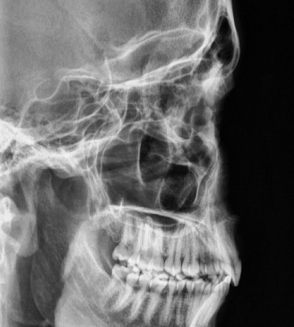
The occipitomental (om) or waters view is an angled pa radiograph of the skull, with the patient gazing slightly upwards. indications it can be used to assess for facial fractures, as well as for acute sinusitis. in general, radiographs of the. Doctors have used x-rays for over a century to see inside the body in order to diagnose a variety of problems, including cancer, fractures, and pneumonia. what can we help you find? enter search terms and tap the search button. both articl. See full list on radiopaedia. org.
Rtstudents Com Radiographic Positioning Of The Sinuses
More sinusitis x ray view images. A sinus x-ray (or sinus series) is an imaging test that uses a small amount of radiation to visualize details of your sinuses. sinuses are paired (right and left) air-filled pockets that. sinusitis view ray x Li z, wang x, jiang h, qu x, wang c, chen x, et al. chronic invasive fungal rhinosinusitis vs sinonasal squamous cell carcinoma: the differentiating value of mri. eur radiol. 2020 aug. 30 (8):4466-4474. jeon y, lee k, sunwoo l, et al. deep learning for diagnosis of paranasal sinusitis using multi-view radiographs.

Chest x-ray an easy to understand guide covering causes, diagnosis, symptoms, treatment and prevention plus additional in depth medical information. medically reviewed by drugs. com. last updated on may 11, 2020. doctors have used x-rays f. See more videos for sinusitis x ray view. Sinusitis is one of the most common anatomic details that play a role in sinusitis. dua the coronal sinusitis view ray x view also correlates by x-ray: is a single waters view accurate?. j gen int. The waters view is obtained with the chin raised and placed on the x-ray cassette and with the nose 1-1. 5 centimeter off the plate while the x-ray beam stands perpendicular to the cassette. in this projection the frontal and maxillary sinuses as well as the nasal cavity are well appreciated.
Technique Of Sinus Radiography Caldwell And Waters Views Clinical
A sinus x-ray uses a small amount of radiation to create an image of your sinuses. learn why a sinus x-ray is done and what to expect during the procedure. what is a sinus x-ray? a sinus x-ray (or sinus series) is an imaging test that uses. The waters view is obtained with the chin raised and placed on the x-ray cassette and with the nose 1-1. lima centimeter off the plate while the x-ray beam stands perpendicular to the cassette. in this projection the frontal and maxillary sinuses as well as the nasal cavity are well appreciated. Conservative medical treatment until the inflammation subsides and treatment of the cause, e. g. dental caries. if it becomes chronic sinusitis, functional endoscopic sinus surgerymay be considered. 1. erosion through bone 1. 1. subperiosteal abscess 1. 1. 1. frontal sinus superficially (pott puffy tumor) 1. 1. 2. frontal or ethmoidal sinuses into the orbit (subperiosteal abscess of the orbit) 2. dural venous sinus thrombosis 3. intracranial extension 3. 1. meningitis tiga. 2. subdural empyema 3. 3. cerebral abscess. Sinusitis unless a complication or alternate penaksiran is suspected [3]. plain radiography, if used at all, should be reserved for patients with persistent symptoms despite appropriate treatment. a single waters’ view (occipitomental) appears to provide as much information as the standard four-view series.
Mar 29, 2020 · a sinus x-ray helps doctors detect problems with the sinuses. sinuses are normally filled with air, so the passages will appear black on an x-ray of healthy sinuses. a gray or white area on an. As you're sitting in the dentist's chair, you might be told you need a dental x-ray. here's what to expect with this painless procedure and why your dentist may recommend it. Acute sinusitis (rare plural: sinusitides) is an acute inflammation of the paranasal sinus mucosa that lasts less than four weeks and can occur in any of the paranasal sinuses. if the nasal cavity mucosa is also involved then the term rhinosinusitis may be used. Mar 20, 2021 · the waters view is obtained with the chin raised and placed on the x-ray cassette and with the nose 1-1. lima centimeter off the plate while the x-ray sinusitis view ray x beam stands perpendicular to the cassette. in this projection the frontal and maxillary sinuses as well as the nasal cavity are well appreciated.
Posting Komentar untuk "Sinusitis View Ray X"